Here are several large animal studies we’ve undertaken to illustrate how mitochondrial transplantation can reduce ischemia-reperfusion injuries and help preserve function of the heart, lungs, and limbs.

Donor after cardiac death (DCD) heart transplantation

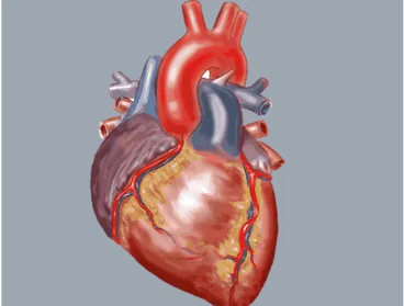
Heart organ

Mitochondrial transplantation for myocardial protection in ex-situ‒perfused hearts donated after circulatory death

Donation after death is limited because of the effects of ischemia. With this line of research, we saw that mitochondrial transplantation after the circulatory death of pig hearts improves their recovery after ischemia and perfusion. The study indicates that mitochondrial transplantation may help preserve function in these types of hearts and expand the neonatal, pediatric and adult donor heart pool.
Myocardial protection in pre- and post-ischemic and diabetic hearts

Heart organ

Mitochondrial transplantation for myocardial protection in diabetic hearts

Type 2 diabetes can lead to mitochondrial dysfunction and increase the risk of heart muscle damage caused by ischemia-reperfusion injury (interrupted and restored blood flow). We have found that the transplantation of mitochondria significantly improves post-ischemic myocardial muscle function recovery and significantly decreases myocellular injury in diabetic hearts.

Autogenous mitochondria transplantation for treatment of right heart failure

Mitochondrial dysfunction is a key event in triggering right ventricular hypertrophy and failure. We found that the injection of autologous mitochondria from healthy muscle preserved heart function and reduced heart muscle cell death, compared with untreated animals. Our findings suggest that mitochondrial transplantation can prevent the loss of heart muscle cells and preserve the pumping function of the right ventricle.

A novel biological strategy for myocardial protection by intracoronary delivery of mitochondria

Using pig models, we discovered that transplanting a patient’s own healthy mitochondria can be an effective and safe therapy for heart muscle ischemia-reperfusion injury. Performing an intracoronary delivery, the therapy increased coronary blood flow and improved myocardial function and tissue perfusion.

Delayed transplantation of autologous mitochondria for cardio-protection in a porcine model

Building upon the pig model study, we wanted to see if delayed mitochondrial transplantation could protect heart muscle from ischemia-reperfusion injury. Again performing intracoronary delivery in pig models, delayed mitochondrial transplantation improved ventricular performance after ischemia and perfusion, suggesting another potential therapeutic strategy for treating interrupted and restored blood flow.

Mitochondrial transplantation prolongs cold ischemia time in murine heart transplantation

Cold ischemia time (CIT) is a critical element of the organ transplantation process; the risk of organ damage increases with every added hour of CIT. With a mouse heart transplant model, we looked to see if mitochondrial transplantation could enhance graft function and viability after prolonged CIT. Indeed, mitochondria injection extended CIT for up to 29 hours in the mouse model, and it significantly improved graft function and decreased graft tissue injury.

Acute kidney injury

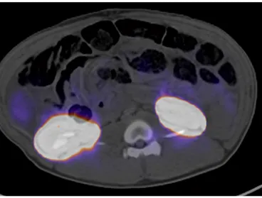
scan of kidneys

A large animal model for acute kidney injury by temporary bilateral renal artery occlusion

Ischemia-reperfusion injury is the most common cause of acute kidney injury. To better understand how the injury could affect human kidneys, our lab recreated the circumstances in a pig model. The approach closely mimicked perioperative renal hypoperfusion, leading us to believe it may improve translation of acute kidney injury research to human clinical settings.

Acute lung injury

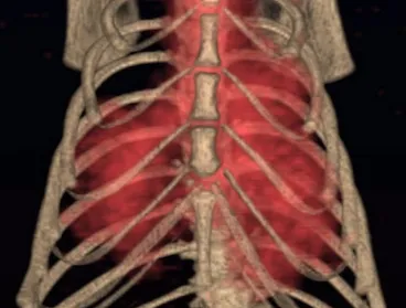
Human ribcage

Mitochondrial transplantation enhances murine lung viability and recovery after ischemia-reperfusion injury

Ischemia-reperfusion injury is also the most common cause of acute lung injury. In a mouse model of lung ischemia-reperfusion injury, mitochondrial transplantation — delivered through the pulmonary artery or by nebulization — was shown to improve lung function and reduced tissue damage. The therapy helped restore near-normal lung mechanics after injury, potentially opening a door to treatment for acute lung injury related to ischemia-reperfusion injury.

Mitochondrial transplantation improves lung function during ex vivo lung perfusion

Using a pig model, isolated mitochondria were delivered through the pulmonary artery during ex vivo perfusion of donor lungs. Mitochondrial transplantation improved lung function and reduced ischemia-reperfusion injury. This approach may provide a new strategy for treating and improving donor organs outside the body before transplantation.

Acute limb ischemia

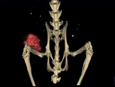
Lower half of human spine and hip bones

Mitochondrial transplantation ameliorates acute limb ischemia

Mitochondrial injury plays a key role in the ischemia-reperfusion injury of skeletal muscle tissue. To further demonstrate that mitochondrial transplantation is an effective therapy to replace or augment damaged mitochondria, we looked at transplanting mitochondria in a mouse acute limb ischemia model. Mice receiving mitochondria showed significantly less tissue and cell death and improved hind-limb function than control mice — suggesting that mitochondrial transplantation protects skeletal muscle and improves limb function following acute limb ischemia.