Mitochondrial Isolation (Boston Children's Hospital)
1. Stock Solutions
0.5 M K+-EGTA
Ethylene glycol-bis(2-aminoethylether)-N,N,N′,N′-tetraacetic acid; Sigma-Aldrich E3889, MW= 380.35
Note: This is an acetic acid.
For 300 mL final
57.05 g EGTA
Bring to approximately 200 mL with dd H2O
Start stirring
Add in solid KOH crystals (approximately 15 grams) watch pH- do not dump in all 15 grams this is just an approximation
EGTA will not go into solution until it is at approximately pH 8.0
When you get to approximately pH 7.6 adjust pH to 8.0 using 5 M KOH solution.
Do not go beyond pH 8.0
Bring to 300 ml with dd H2O
Filter sterilize with 0.01 um filtration unit.
Store at 4oC
1 M K+-HEPES pH 7.2
N-(2-Hydroxyethyl)piperazine-N′-(2-ethanesulfonic acid), 4-(2-Hydroxyethyl) piper-azine-1-ethanesulfonic acid; Sigma-Aldrich H3375
MW 238.3
Note: This is a sulfonic acid.
For 300 mL final
71.49 g HEPES
Bring to approximately 200 mL with dd H2O
Adjust pH with 5 M and then 1 M KOH to pH 7.2
Bring to 300 ml with dd H2O
Filter sterilize with 0.01 um filtration unit.
Store at 4oC
2. Solution A
Final Concentration
| 300 mM Sucrose | 51.34 G |
| 10 mM K+-HEPES pH 7.2 | 5 mL of 1 M K+-HEPES pH 7.2 |
| 1 mM K+-EGTA, pH 8.0 | 1 mL of 0.5 M K+-EGTA pH 8.0 |
| Bring to 500 mL with dd H20 | 500 mL |
Filter sterilize with 0.22 um filtration unit.
Store at 4oC
3. Mitochondrial Isolation Method
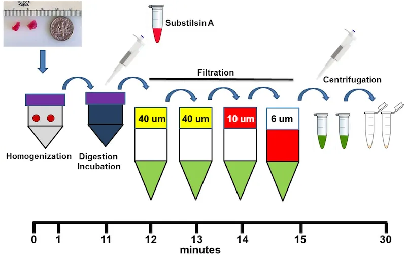
Schematic overview of the rapid isolation of mitochondria from tissue. A small tissue sample is homogenized, enzymatically treated with Subtilisin A, and sequentially filtered through 40-, 40-, 10-, and 6-µm filters to remove tissue debris. The filtrate is then centrifuged to collect the mitochondria. The complete isolation procedure is performed within approximately 30 minutes.
Place sterile Solution A at 4oC.
Prepare Solution B at 4oC. (2 mg Subtilisin A in 1 mL solution A).
Note: Pre-weigh 2 mg Subtilisin A as a dry powder in a 1.5 mL Eppendorf tube and store at -20oC - prior to use we add 1 mL of solution A. Subtilisin A -P5380 Sigma, Protease from Bacillus licheniformis, Type VIII, lyophilized powder, 7-15 units/mg solid
Pre-assemble two 40 um, one 10 um and one 6 m filters in separate sterile 50 mL Falcon tubes and keep at 4oC.
Pre-wet all filters with Solution A – Remember to remove all Solution A from the Falcon tubes prior to filtration so as not to dilute your sample.
Get tissue from fresh source. Use #6 biopsy punch to get tissue pieces. For skeletal muscle use 2 punch samples. For liver use one punch.
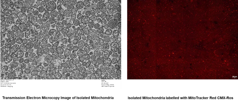
Note: Do not overload the method. The mitochondria you get from this amount of tissue are very pure (see transmission electron microscopy image at the end of this pdf). More tissue does not increase the number but does clog the system. If you need more than 1 x 10¹⁰ mitochondria-do two or more parallel isolations.
Place tissue and 5 mL of Solution A in Miltenyi C tube.
Homogenize the tissue in a Miltenyi homogenizer. Use program h-mitochondria isolation C Tube.
Add Solution B to the homogenate and incubate at 4oC for 10 minutes.
OPTIONAL: Following 10 min. incubation, centrifuge at 750 x G for 4 min.
Note: We don’t usually do this unless there is a lot of suspended debris. Usually after the 10 min. incubation the debris settles to the bottom -don’t mix the contents -you can pour the supernatant directly onto the filter - or use a pipettor- leave the debris in the bottom of the tube.
Following 10 min. incubation, filter the supernatant through the pre-wetted filter unit consisting of a 40 uM cell strainer (BD Falcon REF 352340) in a sterile 50 mL Falcon tube at 4oC.
Note: Use Falcon Brand tubes -the generic tubes are too tight on the filters, and you will get a vacuum.
Pass the collected filtrate though the second pre-wetted filter unit consisting of a 40 uM cell strainer (BD Falcon REF 352340) in a separate, sterile 50 mL Falcon tube at 4oC.
Pass the collected filtrate through the filter unit consisting of a 10 uM filter (Pluri-select cell strainer 10 um, 43-500010-03) in a separate, sterile 50 mL Falcon tube at 4oC.
Pass the collected filtrate through the filter unit consisting of a 6 uM filter (Pluri-select cell strainer 6 um, 43-500006-03) in a separate, sterile 50 mL Falcon tube at 4oC.
Note: This step is optional as we have found that sometimes these 6 uM filters do not work very well and water won’t pass through –we check by pre-wetting to see if they allow flow -I try wetting the top and the bottom to check for flow.
The filtrate is transferred to sterile 1.5 centrifuge tubes (usually -2-3 tubes)
The mitochondria are collected by centrifugation at 9500 RPM in an Eppendorf Centrifuge at 4oC for 5 min.
The supernatant is removed and the mitochondrial pellets are resuspended by triturated by gentle pipetting up and down in 0.5 mL Solution A and pooled at 4oC.
Note: We normally pool all tubes in a total volume of 1 mL Solution A.
Centrifuge at max in Eppendorf centrifuge for 2 min. Discard supernatant.
The mitochondrial pellet is resuspended by trituration, by gentle pipetting up and down in 0.5 mL Solution A.
Re-centrifuge in Eppendorf centrifuge for 2 min. Discard supernatant.
The mitochondrial pellet is resuspended by trituration, by gentle pipetting up and down in 0.5 mL Solution A.
Re-centrifuge in Eppendorf centrifuge for 2 min. Discard supernatant.
Resuspend the mitochondrial pellet by trituration at desired concentrations in Solution A in a 1.5 mL Eppendorf tube.
We use the mitochondria immediately for transplantation.
4. Mitochondria number and Viability
The mitochondria should undergo visual inspection and a cell count with and membrane-potential viability stain (MitoTracker Red CMXRos; Thermo Fisher Scientific),
A: Coulter Counter
Count the isolated mitochondria by particle counter or hemocytometer. The average yield is 3-5 x 10⁹ / mL.
B: Hemocytometer
Mitotracker Red CMXRos is dissolved in 470 uL of fresh DMSO (Sigma Aldrich D2650-5X5ML), approximately 200 uM final concentration and stored at -20oC in aliquots (50-100 uL). Defrost just prior to use.Note: use only fresh unopened DMSO.
Viable mitochondrial number is determined by labeling an aliquot (100 uL) of isolated mitochondria with 5 umol/L MitoTracker Red CMTMRos (Invitrogen, Carlsbad, CA) in a 1.5 mL Eppendorf tube. Cover the tube with foil and incubate on ice for 5 min. We use 2.5 uL of stock. Remove 2 uL of labeled mitochondria and add 18 uL of Respiration Buffer. Mix by trituration (gentle pipetting up and down). The dilution factor is 1:10.
Clean the hemocytometer and cover slip thoroughly. Check cleanliness under the microscope.
Place the hemocytometer and cover slip on the microscope stage.
Place a pipette filled with a well-suspended mix of mitochondria at the notch at the edge of the hemocytometer and then slowly expel 10 uL so that the fluid is drawn into the chamber by capillary action. Repeat on the other side of the hemocytometer.
Count the number of mitochondria in each of the 4 outside large squares. Average the 4 squares. Note that the volume under each square is 0.1 mL x 10-3 and a dilution factor for the hemocytometer of 104 must be used in the calculation.
The mitochondria concentration is calculated as follows: Mitochondrial concentration per mL = (Total cell count /4 ) x 104 (hemocytometer dilution factor) x dilution factor of mitochondrial sample. Example: 500 mitochondria per square x 104 x 10 = 5 x 1 07 mitochondria/mL
Viability can also be determined by FACs analysis using MitoTracker Red CMTMRos (Invitrogen, Carlsbad, CA). Label mitochondria as described above.
- Endotoxin testing is performed by kinetic Limulus amebocyte lysate assay (Endosafe nexgen-PTS; Charles River Laboratories), and Gram stain, and was released after meeting all criteria.
- We use the mitochondria immediately for transplantation.
- ATP content can be determined by the ATPlite Luminescence ATP Detection Assay System (Perkin Elmer, Waltham, MA, USA) (see reference [1] for methodology)
- Compendial sterility testing (USP <71>) of a retention aliquot should be performed.
5. Mitochondria Isolation from Cell Culture
The example used here is for human cardiac fibroblasts that we also use for uptake analysis.
Human adult cardiac fibroblasts (ScienCell, Carlsbad, CA) are maintained in Fibroblast Medium-2 containing fetal bovine serum, fibroblast growth supplement-2, and antibiotic (penicillin/streptomycin) solution according to the supplier’s directions.
Human Cardiac Fibroblast (HCF) Medium Preperation Protocol
Step 1
Human Cardiac Fibroblast (HCF) cells were purchased from ScienCell.(Cat No:6300)
Recommended Medium: Fibroblast Medium-2 (FM-2, ScienCell. Cat: 2331)
Step 2
Complete medium:
500 ml FM-2 (ScienCell. Cat No:6300)
25 ml Fetal Bovine Serum (ScienCell. Cat No:0025)
5 ml of Fibroblast Growth Supplement-2 (FGS) (ScienCell. Cat No: 2382)
5 ml Penicillin/Streptomycin solution (P/S) (ScienCell. Cat No:0503)
Step 3
After adding all supplements, shake the media gently and make sure that the media is mixed homogeneously.
Two 100 mm culture plates of human adult cardiac fibroblasts are grown to approximately 80% confluency.
Remove the media.
Rinse each plate of cells with PBS (pre-warmed to 37⁰C) two times.
Remove the PBS.
Add 0.25% trypsin (pre-warmed to 37⁰C) to the cells (0.5 -1.0 mL).
Incubate in the incubator 1-3 min.
Check the release of cells by microscopy.
Stop trypsinization by adding 5 mL of media containing FBS.
Collect the cells by centrifugation at 1500 x g for 10 min.
Remove the media.
Wash the cell pellet with prewarmed DPBS by centrifugation at 1500 x g for 5 min, and remove the DPBS.
Resuspend the cell pellet in 5 mL of solution A.
Mitochondria are isolated as described in 3.
6. Cell Culture Coincubation - uptake and ATP content
Have cells, media and all cell culture equipment ready for co-incubation. We have found that 50,000 cells/well in a 24-well plate is optimal. We culture the cells overnight and perform experiments the next morning.
Isolate the human mitochondria from cells as described in 5 o5 from from tissue as described in 5 and 3.
Label the isolated mitochondria using 2 uL of MitoTracker Red CMXRos per 1000 ul. Incubate in the dark at 37oC for 5 min.
MitoTracker Red CMXRos is dissolved in 470 uL of fresh DMSO (Sigma Aldrich D2650-5X5ML), approximately 200 uM final concentration, and stored at -20oC.
Note: To allow for simultaneous estimation of mitochondrial uptake into cells and ATP content, we usually perform these experiments in parallel with duplicate samples using labeled mitochondria for uptake and unlabeled mitochondria for ATP lite assay.
Note: If you are using the isolated mitochondria for co-incubation with cell culture, it is important to wash the isolated mitochondrial pellet 4 times in Solution A.
Wash the non-labeled and the labelled mitochondria as described below.
Centrifuge at max in Eppendorf centrifuge for 2 min. Discard supernatant. The mitochondrial pellet is resuspended by trituration, by gentle pipetting up and down in 0.5 mL Solution A.
Re-centrifuge in an Eppendorf centrifuge for 2 min. Discard supernatant. The mitochondrial pellet is resuspended by trituration, by gentle pipetting up and down in 0.5 mL Solution A.
Re-centrifuge in an Eppendorf centrifuge for 2 min. Discard supernatant. The mitochondrial pellet is resuspended by trituration, by gentle pipetting up and down in 0.5 mL Solution A.
Re centrifuge in Eppendorf centrifuge for 2 min.
Save supernatant from this last wash for negative control.
The mitochondrial pellet is resuspended by trituration, by gentle pipetting up and down at desired concentrations in Solution A in a 1.5 mL Eppendorf tube.
We use the mitochondria immediately for transplantation.
Mitochondria number for co-incubation can vary; however, we have found that concentrations less than or equal to 1 x 10⁷ / 50,000 cells in a 24-well plate are optimal.
Co-Incubation Method
Remove the media from each well on the cell plate to be used for co-incubation.
Add 100-200 uL of media containing mitochondria at the desired concentration in each well.
Note: Mitochondria float, as evidenced by differential centrifugation techniques for isolation and thus for co-incubation to occur the mitochondria must be in contact with the cell- use a minimal volume to just cover the cells.
Negative control cells are incubated with 100-200 uL media containing the saved last wash.
Incubate cells as usual for the required time. If incubating beyond 24 hours, add fresh media at 24 hours as appropriate.
Determine uptake by fluorescence microscopy.
7. Slide Chamber Fluorescence Microscope Method
Material:
Poly-L-lysine solution (PLL, 0.1 % (w/v) in H2O)- (Sigma)-P8920
Thermo Scientific™ Nunc™ Lab-Tek™ II Chamber Slide™ System- 154526
MitoTracker Green FM – Invitrogen- M7514
MitoTracker Red CMXRos- Invitrogen- M7512
DAPI (Thermo Fisher - D1306)
DPBS
Coat the slide surface with 1 mL of 0.1% Poly-L-lysine solution (PLL, 5 mg in 50 mL H2O).
Incubate for 5 min. Then remove the PLL by aspiration.
Rinse the surface 4 times with sterile ddH2O and aspirate the washes.
Allow the slide to dry overnight.
The cells to be transplanted with mitochondria are seeded at 50,000 per chamber and grown overnight. Ensure the cells are fully attached.
Isolate mitochondria as described in 3.
Label the isolated mitochondria using 2 uL of MitoTracker Red CMXRos per 1000 ul. Incubate in dark at 37oC for 5 min.
MitoTracker Red CMXRos is dissolved in 470 uL of fresh DMSO (Sigma-Aldrich D2650-5X5ML), approximately 200 uM final concentration, and stored at -20oC).
Wash the labeled mitochondria as described below:
Centrifuge at max in an Eppendorf centrifuge for 2 min. Discard supernatant.
The mitochondrial pellet by trituration, by gentle pipetting up and down in 0.5 mL Solution A.
Re-centrifuge in an Eppendorf centrifuge for 2 min. Discard supernatant.
The mitochondrial pellet is resuspended by trituration, by gentle pipetting up and down in 0.5 mL Solution A.
Re-centrifuge in an Eppendorf centrifuge for 2 min. Discard supernatant.
The mitochondrial pellet is resuspended by trituration, by gentle pipetting up and down in 0.5 mL Solution A.
Re-centrifuge in an Eppendorf centrifuge for 2 min. Save supernatant from this last wash for negative control.
The mitochondrial pellet is resuspended by trituration, by gentle pipetting up and down .at desired concentration in Solution A in a 1.5 mL Eppendorf tube.
While labeling the isolated mitochondria, label the cells in the slide chamber with 200 uL of media containing 1 uL MitoTracker Green FM for 30 min. in the dark at 37oC.
MitoTracker Green FM is dissolved in 372 uL of fresh DMSO (Sigma Aldrich D2650-5X5ML), approximately 200 uM final concentration and stored at -20oC).
The cells labeled with MitoTracker Green FM are washed twice with warm PBS (1X).
The PBS is removed and media containing mitochondria is added.
Mitochondria labeled with MitoTracker Red CMXRos are suspended in the media and added to each well at 200 µl per well.
Mitochondria number for co-incubation can vary, however, we have found that concentrations less than or equal to 1 x 10⁷ / 50,000 cells in a 24-well plate are optimal.
Add 100-200 uL of media containing mitochondria at desired concentration in each well.
Note: Mitochondria float, as evidenced by differential centrifugation techniques for isolation and thus for co-incubation to occur the mitochondria must be in contact with the cell- use a minimal volume to just cover the cells.
Control cells are incubated with 100-200 uL media containing the saved last wash.
Incubate cells as usual for required time. If incubating beyond 24 hours add fresh media at 24 hours as appropriate.
For DAPI staining-the media is discarded and the well is washed 2 times with warm DPBS (1X) and stained with DAPI for 5 minutes in the dark at 37oC.
DAPI stock is made by adding 2 ml H2O to the DAPI dry contents (Thermo-Fisher -D1306).
Add 2.1 uL of stock to 100 ul 1X PBS. For staining cells, use 1:1000 in media.
Following 5 min incubation, remove the DAPI media and wash the cells 3 times in DPBS.
Determine uptake by fluorescence microscopy.
8. Determining uptake using human mitochondria
In our experimental studies we use unlabeled autologous mitochondria, however for uptake studies, we have found that the use of human mitochondria provides for unambiguous results.
The use of human mitochondria in an animal model allows for the differentiation between endogenous animal mitochondria and transplanted human mitochondria based on immune reactivity to a monoclonal anti-human mitochondria antibody (Cowan et al., 2017).
When used with histology analysis this methodology allows for identification of mitochondria based on shape and immune reactivity and allows for localization with other cellular elements. Alternative methods using fluorescent labels or polymerase chain reaction (PCR) analysis to estimate uptake while useful are not specific as they could reflect either transfer of label or identification of mitochondrial constituents and not intact mitochondria.
We use human cardiac fibroblasts as our human mitochondrial source due to their mitochondrial content; however, any human cell line can be used.
Note: In our initial studies we used HeLa cells for analysis to provide ‘proof of concept’. HeLa cells have few mitochondria and may plates are needed for sufficient mitochondrial isolation.
Cell Culture
As described in 5.
Note: The mitochondria can be labeled with MitoTracker CMXros but the color should not interfere with detection by human mono-clonal antibodies (Alexa 488-phalloidin, green).
The cells are fixed with 4% paraformaldehyde in PBS for 1 hour at 4°C.
The cells are permeabilized for 3 min with 0.1% Triton X-100 in PBS.
Human mitochondria in animal cells are detected using the anti-human mitochondria MTC02 monoclonal antibody (Abcam) at a concentration of 5 ug/ml and detected with Alexa 488-phalloidin (1:50 dilution).
Uptake is determined by fluorescence microscopy.
9. ATP content in mitochondria
We use the ATPlite Luminescence ATP Detection Assay System (Perkin Elmer, Waltham, MA, USA) (see reference [1] for methodology).
10. Oxygen Consumption Analysis
If you are going to do Oxygen consumption – note that the mitochondria isolated by this method are energized.
11. Transmission Electron Microscopy Analysis
Isolate mitochondria as described in 3.
Collect the mitochondrial pellets as described in 3.
Combine the pellets by centrifugation.
Remove solution A.
Resuspend the combined pellet in fixative.
Fixative: Routine fixative; 2.5% Glutaraldehyde 1.25% Paraformaldehyde and 0.03% picric acid in 0.1 M sodium cacodylate buffer (pH 7.4)
The fixative is provided to us as a 2x of the above solution (5%Glut, 2.5% PFA, 0.06% picric acid in 0.2M cacodylate buffer pH 7.4).
We add the 2x solution 1:1 with our mitochondria or cells and spin down immediately. Leave the mitochondria, cells to fix as a pellet for at least 2 hours at RT or ON at 4C.
*WE BRING THE PELLET TO THE EM LAB IN THE FIXATIVE.
The description of the method is as follows:
A pellet of cells was fixed for at least 2 hours at RT in the above fixative, washed in 0.1M cacodylate buffer and postfixed with 1% Osmiumtetroxide (OsO4)/1.5% Potassiumferrocyanide(KFeCN6) for 1 hour, washed 2x in water, 1x Maleate buffer (MB) 1x and incubated in 1% uranyl acetate in MB for 1hr followed by 2 washes in water and subsequent dehydration in grades of alcohol (10min each; 50%, 70%, 90%, 2x10min 100%). The samples were then put in propylene oxide for 1 hr and infiltrated ON in a 1:1 mixture of propylene oxide and TAAB Epon (Marivac Canada Inc. St. Laurent, Canada). The following day, the samples were embedded in TAAB Epon and polymerized at 60 degrees C for 48 hrs.
Ultrathin sections (about 60nm) were cut on a Reichert Ultracut-S microtome, picked up onto copper grids stained with lead citrate, and examined in a JEOL 1200EX Transmission electron microscope or a TecnaiG² Spirit BioTWIN, and images were recorded with an AMT 2k CCD camera.
References:
Preble JM, Pacak CA, Kondo H, McKay AA, Cowan DB, McCully JD. Rapid Isolation and Purification of Mitochondria for Transplantation. J Vis Exp. 2014; 91: e51682. doi: 10.3791/51682. PMID: 25225817
Preble JM, Kondo H, Levitsky S, James D. McCully JD. Quality Control Parameters for Mitochondria Transplant in Cardiac Tissue. JSM Biochem Mol Biol 2014 2(1): 1008
12. Fluorescent Staining of Formalin-Fixed, Paraffin-Embedded sections
STEP | DESCRIPTION | SOLUTION | PLACE | TEMP | TIME | COMMENTS |
|---|---|---|---|---|---|---|
1 | XYLENE | Xylene | Fume Hood | RT | 5 min X 2 | |
2 | RE-HYDRATION | 100% Ethanol | Bench | RT | 5 min | |
90% Ethanol | Bench | RT | 5 min | |||
80% Ethanol | Bench | RT | 5 min | |||
50% Ethanol | Bench | RT | 3 min | |||
3 | WASH | PBS | Bench | RT | 3-5 min | |
4 | ANTIGEN RETRIEVAL | AR Buffer | Bench | 90-95°C | 30 min | watch temp carefully, in 500ml glass container |
5 | COOL DOWN | AR Buffer | Cold Water Bath | ~10 min | ||
6 | WASH | PBS | Bench | RT | 3 min | |
Mark slides with Super PAP Pen (liquid repellent) | ||||||
7 | BLOCKING | 5% horse serum | Bench | RT | 30 min | |
8 | 1ry ANTIBODY | 1ry Ab in blocking buffer | Bench | RT | overnight | |
9 | WASH | PBS | Bench | RT | 3 min X 3 | |
10 | 2ry ANTIBODY (Fluorescent) | 2ry Ab in blocking buffer | Bench, Dark | RT | 1 hour | |
11 | WASH | PBS | Bench, Dark | RT | 3 min X 3 | |
12 | DAPI Staining | DAPI in PBS | Bench, Dark | RT | 10 min | |
13 | WASH | PBS | Bench | RT | 3 min X 3 | |
14 | WASH | dH2O | Bench | RT | rinse | |
15 | MOUNTING | Dako Fluorescence Mounting Medium | Bench | RT | - | Stored at 4oC, DARK |
Primary Ab: 1:100 dilution (200uL per slide)
Secondary Ab: 1:200 dilution (200uL per slide)
Materials
- Distilled water
- PBS
- Xylene
- Ethanol
- Hydrochloric Acid
- Trizma base (Sigma, T6791)
- 0.5M EDTA, pH 8.0 (Invitrogen, 15575-020)
- Tween-20 (Bio-Rad, 170-6531)
- Horse serum (Vector Laboratory)
- DAPI (Invitrogen; D1306)
Antigen Retrieval
For paraffin-embedded sections, I dewax in xylene 3 times and rehydrate through a graded ethanol series (100%, 95%, 95%, 70%, 70%, 50%, water, PBS). I then boil the slides in a 1200W microwave 3 time for 5 min each in 1 mM EDTA, pH 8.0.
- Slides were baked for 1 hour at 65°C,
- Deparaffinize sections in xylene, 3 x 5 min.
- Hydrate with 100% ethanol, 2 x 3min.
- Hydrate with 95% ethanol, 1min.
- Hydrate with 95% ethanol, 1min
- Hydrate with 70% ethanol, 1min
- Hydrate with 70% ethanol, 1min
- Hydrate with 50% ethanol, 1min
- Rinse in distilled water.
- Rinse in PBS.
Antigen Retrieval
- Put 1 mM EDTA, pH 8.0 in a microwave container to cover the sides and top of the slides
- Boil the slides in a 1200W microwave 3 times for 5 min each in 1 mM EDTA, pH 8.0.
- Change buffer each time
- Cool on the bench
Reference:
Cowan DB, Yao R, Akurathi V, Snay ER, Thedsanamoorthy JK, Zurakowski D, Ericsson M, Friehs I, Wu Y, Levitsky S, Del Nido PJ, Packard AB, McCully JD. Intracoronary Delivery of Mitochondria to the Ischemic Heart for Cardioprotection. PLoS One. 2016 Aug 8;11(8):e0160889. doi: 10.1371/journal.pone.0160889. PMID: 27500955; PMCID: PMC4976938.